Anatomy Of Chest / Female Anatomy Of Chest And Abdomen On White Background 2 Stock Illustration Adobe Stock / The circulatory system does most of its.. Anatomical quadrants 12 photos of the anatomical quadrants 9 anatomical quadrants, anatomical quadrants and regions, anatomical quadrants of the abdomen, anatomical quadrants of the body, four abdominal quadrants, human anatomy, 9 anatomical quadrants, anatomical quadrants and regions, anatomical quadrants of the abdomen. The right side of the heart is deflected anteriorly, and the left side is deflected posteriorly. Find out more about the individual muscles within the chest anatomy by clicking their respective. See chest anatomy stock video clips. See human chest anatomy stock video clips.
Sternocleidomastoid muscle clavicle and ribs anatomy muscle anatomy chest sternocleidomastoid ribs anatomy chest muscles anatomy thorax rib muscles chest muscles chest anatomy illustration. Learn about each of these muscles, their locations, functional anatomy and exercises for them. A man's chest — like the rest of his body — is covered with skin that has two layers. Thoracic cavity, also called chest cavity, the second largest hollow space of the body.it is enclosed by the ribs, the vertebral column, and the sternum, or breastbone, and is separated from the abdominal cavity (the body's largest hollow space) by a muscular and membranous partition, the diaphragm.it contains the lungs, the middle and lower airways—the tracheobronchial tree—the heart. This page provides an overview of the chest muscle group.
Imaging Anatomy Chest Abdomen Pelvis Von Michael P Federle Isbn 978 0 323 47781 9 Fachbuch Online Kaufen Lehmanns De from www.lehmanns.de Computed tomography (ct) of the chest can detect pathology that may not show up on a conventional chest radiograph (1). Mar 24, 2021 · related posts of anatomy of the chest area anatomical quadrants. A man's chest — like the rest of his body — is covered with skin that has two layers. The chest or thorax is the region between the neck and diaphragm that encloses organs, such as the heart, lungs, esophagus, trachea, and thoracic diaphragm. Learn about each of these muscles, their locations, functional anatomy and exercises for them. This page provides an overview of the chest muscle group. The circulatory system does most of its. It is important to remember the position and orientation of the heart when placing a stethoscope on the chest of a patient and listening for heart sounds, and also when looking at images taken from a midsagittal perspective.
Thoracic cavity, also called chest cavity, the second largest hollow space of the body.it is enclosed by the ribs, the vertebral column, and the sternum, or breastbone, and is separated from the abdominal cavity (the body's largest hollow space) by a muscular and membranous partition, the diaphragm.it contains the lungs, the middle and lower airways—the tracheobronchial tree—the heart.
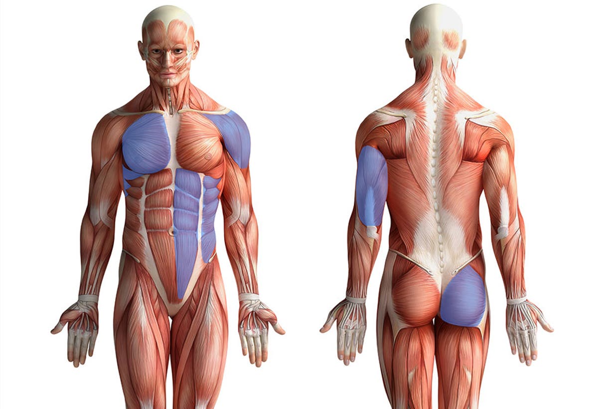
Sep 14, 2015 · the chest anatomy includes the pectoralis major, pectoralis minor and the serratus anterior. The chest or thorax is the region between the neck and diaphragm that encloses organs, such as the heart, lungs, esophagus, trachea, and thoracic diaphragm. Sternocleidomastoid muscle clavicle and ribs anatomy muscle anatomy chest sternocleidomastoid ribs anatomy chest muscles anatomy thorax rib muscles chest muscles chest anatomy illustration. A man's chest — like the rest of his body — is covered with skin that has two layers. Mar 24, 2021 · related posts of anatomy of the chest area anatomical quadrants. See chest anatomy stock video clips. The circulatory system does most of its. This page provides an overview of the chest muscle group. See human chest anatomy stock video clips. Computed tomography (ct) of the chest can detect pathology that may not show up on a conventional chest radiograph (1). Learn about each of these muscles, their locations, functional anatomy and exercises for them. Oct 15, 2017 · radiology basics of chest ct anatomy with annotated coronal images and scrollable axial images to help medical students and junior doctors learning anatomy. Jan 20, 2018 · chest.
Find out more about the individual muscles within the chest anatomy by clicking their respective. The chest or thorax is the region between the neck and diaphragm that encloses organs, such as the heart, lungs, esophagus, trachea, and thoracic diaphragm. The epidermis is the outermost layer that provides a protective, waterproof seal over the body. Anatomical quadrants 12 photos of the anatomical quadrants 9 anatomical quadrants, anatomical quadrants and regions, anatomical quadrants of the abdomen, anatomical quadrants of the body, four abdominal quadrants, human anatomy, 9 anatomical quadrants, anatomical quadrants and regions, anatomical quadrants of the abdomen. It is important to remember the position and orientation of the heart when placing a stethoscope on the chest of a patient and listening for heart sounds, and also when looking at images taken from a midsagittal perspective.
Pushups Way More Than Chest Training from images.contentstack.io The epidermis is the outermost layer that provides a protective, waterproof seal over the body. System respiratory respiratory organs of human body digestive and respiratory system medical chest internal structure of human body medicine body lungs biology intestines stomach anatomy torso human internal. Find out more about the individual muscles within the chest anatomy by clicking their respective. Learn about each of these muscles, their locations, functional anatomy and exercises for them. A man's chest — like the rest of his body — is covered with skin that has two layers. Sep 14, 2015 · the chest anatomy includes the pectoralis major, pectoralis minor and the serratus anterior. Thoracic cavity, also called chest cavity, the second largest hollow space of the body.it is enclosed by the ribs, the vertebral column, and the sternum, or breastbone, and is separated from the abdominal cavity (the body's largest hollow space) by a muscular and membranous partition, the diaphragm.it contains the lungs, the middle and lower airways—the tracheobronchial tree—the heart. Computed tomography (ct) of the chest can detect pathology that may not show up on a conventional chest radiograph (1).
Learn about each of these muscles, their locations, functional anatomy and exercises for them.
See human chest anatomy stock video clips. It is important to remember the position and orientation of the heart when placing a stethoscope on the chest of a patient and listening for heart sounds, and also when looking at images taken from a midsagittal perspective. Computed tomography (ct) of the chest can detect pathology that may not show up on a conventional chest radiograph (1). System respiratory respiratory organs of human body digestive and respiratory system medical chest internal structure of human body medicine body lungs biology intestines stomach anatomy torso human internal. This page provides an overview of the chest muscle group. Thoracic cavity, also called chest cavity, the second largest hollow space of the body.it is enclosed by the ribs, the vertebral column, and the sternum, or breastbone, and is separated from the abdominal cavity (the body's largest hollow space) by a muscular and membranous partition, the diaphragm.it contains the lungs, the middle and lower airways—the tracheobronchial tree—the heart. The right side of the heart is deflected anteriorly, and the left side is deflected posteriorly. The chest or thorax is the region between the neck and diaphragm that encloses organs, such as the heart, lungs, esophagus, trachea, and thoracic diaphragm. Mar 24, 2021 · related posts of anatomy of the chest area anatomical quadrants. The circulatory system does most of its. Jan 20, 2018 · chest. See chest anatomy stock video clips. Mar 18, 2015 · the chest is the area of origin for many of the body's systems as it houses organs such as the heart, esophagus, trachea, lungs, and thoracic diaphragm.
See human chest anatomy stock video clips. Mar 24, 2021 · related posts of anatomy of the chest area anatomical quadrants. The right side of the heart is deflected anteriorly, and the left side is deflected posteriorly. This page provides an overview of the chest muscle group. Mar 18, 2015 · the chest is the area of origin for many of the body's systems as it houses organs such as the heart, esophagus, trachea, lungs, and thoracic diaphragm.
Anatomy Of The Thorax Ct from www.imaios.com Computed tomography (ct) of the chest can detect pathology that may not show up on a conventional chest radiograph (1). System respiratory respiratory organs of human body digestive and respiratory system medical chest internal structure of human body medicine body lungs biology intestines stomach anatomy torso human internal. The epidermis is the outermost layer that provides a protective, waterproof seal over the body. Thoracic cavity, also called chest cavity, the second largest hollow space of the body.it is enclosed by the ribs, the vertebral column, and the sternum, or breastbone, and is separated from the abdominal cavity (the body's largest hollow space) by a muscular and membranous partition, the diaphragm.it contains the lungs, the middle and lower airways—the tracheobronchial tree—the heart. Anatomical quadrants 12 photos of the anatomical quadrants 9 anatomical quadrants, anatomical quadrants and regions, anatomical quadrants of the abdomen, anatomical quadrants of the body, four abdominal quadrants, human anatomy, 9 anatomical quadrants, anatomical quadrants and regions, anatomical quadrants of the abdomen. Mar 24, 2021 · related posts of anatomy of the chest area anatomical quadrants. Sep 14, 2015 · the chest anatomy includes the pectoralis major, pectoralis minor and the serratus anterior. It is important to remember the position and orientation of the heart when placing a stethoscope on the chest of a patient and listening for heart sounds, and also when looking at images taken from a midsagittal perspective.
Thoracic cavity, also called chest cavity, the second largest hollow space of the body.it is enclosed by the ribs, the vertebral column, and the sternum, or breastbone, and is separated from the abdominal cavity (the body's largest hollow space) by a muscular and membranous partition, the diaphragm.it contains the lungs, the middle and lower airways—the tracheobronchial tree—the heart.
Thoracic cavity, also called chest cavity, the second largest hollow space of the body.it is enclosed by the ribs, the vertebral column, and the sternum, or breastbone, and is separated from the abdominal cavity (the body's largest hollow space) by a muscular and membranous partition, the diaphragm.it contains the lungs, the middle and lower airways—the tracheobronchial tree—the heart. Jan 20, 2018 · chest. Oct 15, 2017 · radiology basics of chest ct anatomy with annotated coronal images and scrollable axial images to help medical students and junior doctors learning anatomy. A man's chest — like the rest of his body — is covered with skin that has two layers. Learn about each of these muscles, their locations, functional anatomy and exercises for them. System respiratory respiratory organs of human body digestive and respiratory system medical chest internal structure of human body medicine body lungs biology intestines stomach anatomy torso human internal. The circulatory system does most of its. Mar 24, 2021 · related posts of anatomy of the chest area anatomical quadrants. The epidermis is the outermost layer that provides a protective, waterproof seal over the body. The right side of the heart is deflected anteriorly, and the left side is deflected posteriorly. Sep 14, 2015 · the chest anatomy includes the pectoralis major, pectoralis minor and the serratus anterior. Mar 18, 2015 · the chest is the area of origin for many of the body's systems as it houses organs such as the heart, esophagus, trachea, lungs, and thoracic diaphragm. The chest or thorax is the region between the neck and diaphragm that encloses organs, such as the heart, lungs, esophagus, trachea, and thoracic diaphragm.


0 Komentar